

Rediscovering Biology: Molecular to Global Perspectives
Online Textbook and Video
This online textbook chapter supports and extends the content of the Microbial Diversity video. The chapter covers microbial energy sources, polymerase chain reaction (PCR), and the role of microbes in nutrient cycling. Click on the Go button to begin reading, or skip to a sub-section of the chapter in the list below.
1. Introduction
“If we look we’ll find ’em… the microbes are there. They’re these little packages of secrets that are waiting to be opened.”
– Anna-Louise Reysenbach
Microbes flourish. Inside your gut, in the mucky soil of a marsh, in Antarctic ice, in the hot springs of Yellowstone, in habitats seemingly incompatible with life, microbes flourish.
They were present on Earth 3.5 to 4 billion years ago, and they’ve been evolving and expanding into new environments ever since. Replicating quickly, exchanging genetic material with each other and with other organisms, bacteria and archaea have become ubiquitous.
Not only are they everywhere, but these tiny organisms also manipulate the environments in which they live. Their presence has driven the development of new ecosystems – some of which allowed for the evolution of more complex organisms. Without microbes, the recycling of essential nutrients on Earth would halt. Microbes communicate; some generate the signals for the formation of metabolically diverse communities. Some use sophisticated signaling to establish complex relationships with higher organisms.
In this unit we will examine examples of the broad diversity of microorganisms and consider their roles in various ecosystems, both natural and man-made. We will also discuss some of the practical applications that derive from the wealth of metabolic diversity that microorganisms possess.
Let’s start at the beginning … three or four billion years ago.
2. Microbes as the First Organisms
No one knows for certain where life began. Hot springs and volcanic (hydrothermal) vents on the ocean floor, however, may represent the kinds of environments where cellular life began. Before the ozone layer formed, the surface of Earth was exposed to strong radiation. Thus, most of the Earth’s earliest organisms probably developed beneath the terrestrial surface or in the oceans. It’s likely that these early microbes adapted to the high temperatures associated with abundant volcanic activity. Geological turmoil resulted in the accumulation of carbon dioxide in the atmosphere.
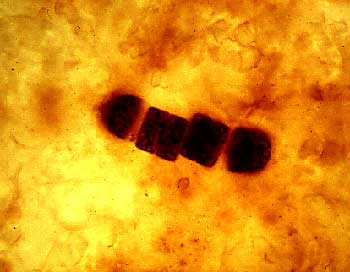
Sometime later, about 2 or 2.5 billion years ago, gaseous oxygen began to appear. Unlike the carbon dioxide, oxygen almost certainly came about because of microbes. Microbes similar to today’s cyanobacteria were present at this time. We know this based on the presence of stromatolites — fossilized microbial mats consisting of layers of filamentous prokaryotes — and trapped sediment that date back to that time. Stromatolite-forming bacteria obtain carbon from carbon dioxide and get their energy by photosynthesis, splitting water to generate oxygen gas in the process. These organisms brought the oxygen level in Earth’s atmosphere to about ten percent of what it is today – enough to allow the evolution of oxygen-using organisms. Gaseous oxygen also contributed to the formation of the ozone layer, which blocks UV radiation. New terrestrial habitats were now open for an evolving diversity of microbes.
3. The Diversity of Microbial Metabolism
The diverse environments on Earth today present energy, and carbon and other nutrients in varying forms. They also vary with respect to temperature, acidity, and the availability of byproducts from other organisms. Microbes thrive in a vast array of these environments.
Microorganisms vary with regard to the sources of energy they use for assembling macromolecules and other cellular components from smaller molecules.

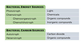
Figure 1. Terms describing varying energy and carbon sources
Phototrophs obtain their energy from light; chemotrophs use chemicals as energy sources. (Troph is derived from a Greek word meaning “to feed.”) Many organisms use organic compounds as sources of energy; these are the chemoorganotrophs. In contrast, the chemolithoautotrophs use inorganic chemicals as energy sources.
Microorganisms also vary with respect to the source of carbon they use. Autotrophs are able to build organic molecules from carbon dioxide. Heterotrophs, the “other feeders,” obtain their carbon from organic compounds – amino acids, fatty acids, sugars, and so on – of autotrophs.
These terms are often combined. So, a “photoautotroph” is an organism that, like plants, gets its energy from light and its carbon from CO2. Decomposers are often chemoheterotrophs; they may obtain energy and carbon from the same source.
So what metabolic classes might microbes found in a deep-sea hydrothermic vent fall within? The lack of sunlight makes them dependent on chemical energy; thus, they are chemotrophs. Carbon dioxide dissolved in the ocean is their source of carbon; they are autotrophs. Organic material from decomposing phototrophs is not abundant, so these organisms rely on inorganic sources for energy. They may use H2 (present in magmatic gases), reduced sulfur compounds or methane as a source of energy. They are also thermophiles, growing optimally at temperatures above 45° C. Thermophilic chemolithoautotrophs serve as primary producers, the first organisms in food chains that include animals such as tube worms and giant clams.
4. Archaea and Bacteria
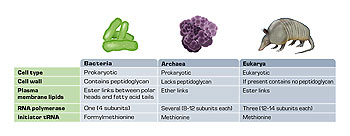
As reviewed in “Evolution and Phylogenetics,” living organisms can be grouped into three domains: the Archaea, the Bacteria, and the Eukarya. Members of Bacteria and Archaea are prokaryotes: single-celled organisms lacking true nuclei and other membrane-enclosed organelles. Bacteria and archaea, however, differ in cell wall characteristics and membrane lipid composition. They also differ in RNA polymerase structure and, therefore, protein synthesis.

Figure 2. Comparison of key characteristics from the three domains of life.
Many extremophiles (organisms that tolerate high or low temperature, high salinity, or extreme pH) fall within the Archaea. Some archaea, the extreme halophiles (salt lovers), tolerate salt concentrations as high as nearly ten times that of seawater.
They have also been found thriving in the Great Salt Lake and the Dead Sea. Nevertheless, habitat alone does not differentiate the groups. Some bacteria grow at temperatures above 80° C, and some Archaea have been found in environments not considered extreme. For example, methanogenic archaea live in anoxic sediments in marshes and are used in sewage treatment facilities. Another archaean, Methanobrevibacter smithii, lives and generates methane in the human colon.
5. The Universal Tree of Life
Starting in the 1970s Carl Woese proposed that variation in the sequences of DNA encoding ribosomal RNA (rRNA) in different organisms would provide valuable information regarding evolutionary relatedness. rRNA is an integral part of ribosomal structure, so it is found in all organisms. After comparing the small variations between the genes for rRNA from many organisms, Woese suggested that the Archaea constitute a unique domain of life, a grouping broader than kingdom. The genomes of several members of the Archaea have been entirely sequenced and have been compared with the genomes of other organisms. Such studies confirm that Archaea constitute a separate group: These organisms contain hundreds of genes with no counterparts in Bacteria or Eukarya. Unexpectedly, ribosomal proteins from Archaea were found to be more similar to those of Eukarya than to Bacterial ribosomal proteins. So, Archaea and Eukarya seem more closely related than Bacteria and Eukarya. (See the Evolution and Phylogenetics unit.)
Can we construct a tree illustrating the relatedness of the three domains, with one common ancestor for all life? Woese and his colleagues have argued — based on phylogenetic methodology and data from several genes — that there is a common ancestor. They further argue that Archaea and Eukarya are more closely related to each other, and that Bacteria diverged from the common ancestor first. (See the Evolution and Phylogenetics unit.)
Other biologists have countered that the true universal tree of life may be more complicated than the picture that Woese and his colleagues presented. The complication is lateral gene transfer, where individuals exchange genes between one another. Although not generally exhibited in Eukarya, mechanisms for lateral gene transfer (also known as horizontal gene transfer) are well known in Bacteria. Genes are exchanged between bacterial species by the action of viruses and by conjugation (cell-to-cell contact in which DNA copied from a plasmid or chromosome is transferred to a recipient cell). Under special conditions, some bacteria are known to take up “naked” DNA from the environment.
Lateral gene transfer, if restricted to very similar organisms, would not pose a problem for constructing a universal tree of life. However, there is evidence that genes have been exchanged between very distant organisms. Eukarya acquired mitochondrial and chloroplast DNA from Bacteria. Nuclear genes in eukaryotes seem to be derived from Bacteria as well, not just from Archaea. Genes are also shared between Archaea and Bacteria.

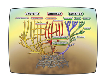
Figure 3. The “Shrub of Life”
Proposed by W. Ford Doolittle, this view of early evolution suggests multiple primitive cells as ancestors to the three domains, and illustrates lateral gene transfer among early organisms.
Twenty-four percent of the genome of the bacterium Thermotoga maritima contains archaen DNA. Similarly, the archaean Archaeoglobus fulgidus has numerous bacterial genes. Some scientists believe that a more diverse community of primitive cells gave rise to the three domains and that the notion of a single universal ancestor might be replaced. W. Ford Doolittle (Dalhousie University) has suggested that lateral gene transfer among early organisms has generated a “tree of life,” which more closely resembles a shrub with untreelike links (shared genes) connecting the branches (Fig. 3).
6. Studying Unculturable Microbes with PCR
Imagine yourself on a team studying archaea at a deep-sea hydrothermal vent at the Galapagos Rift (an area known for its hydrothermal activity). You’ve found a new microbe. What do you want to know about it? What metabolic class does the microbe fall within? Does it make certain proteins? How does it survive the volcanic heat? Traditionally, asking such questions involved growing microbes in the laboratory. Unfortunately, replicating the conditions in which many bacteria and archaea grow is very difficult. For this reason, only a small fraction (perhaps only as few as one percent) of the microorganisms in nature has been cultivated. To identify and compare unculturable organisms microbiologists have turned to molecular genetic techniques.

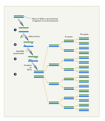
Figure 4. 1) Double-stranded DNA in the sample is heated to generate single strands. 2) Sequence specific primers are added, which anneal to desired sites on the DNA. 3) Nucleotides and heat-tolerant DNA polymerase allow for primer extension at elevated temperature. 4) The result is two new copies of double-stranded DNA. The process is repeated to generate multiple specific dsDNA molecules.
Polymerase chain reaction (PCR) is one technique for studying organisms that cannot be grown in the laboratory. When only a small quantity of DNA is available from a particular source, PCR can be used to amplify that DNA and produce billions of copies of a designated gene-sized fragment. The technique has many applications, including the amplification of DNA from crime scenes, analysis of cancer genes, and identification of pathogens. When an environmental sample contains unculturable organisms, scientists can use PCR to generate copies of microbial genes suitable for comparison.
To replicate DNA in vitro, PCR takes advantage of a special property of the molecule: the hydrogen bonds. These bonds, which bind the complementary strands of DNA together in a double helix, are broken at elevated temperatures (about 95° C). Each single-stranded piece of DNA (ssDNA) is then built upon to form a new, double-stranded molecule (dsDNA). To initiate this, short “primers” — specific ssDNA fragments called oligonucleotides — must anneal to complementary regions on the single-stranded DNA. Deoxynucleotides (A,T,G, and C) and DNA polymerase are added and, in a process called primer extension, the complementary copy of the ssDNA fragment is built. The result is two double-stranded DNA molecules identical to the original. Repeating these steps thirty times can result in a 109-fold amplification of the original molecule.
Careful thermal cycling is required for PCR to proceed. For the primers to anneal to the ssDNA fragments, the temperature is reduced to about 55° C. However, at this temperature the original complementary ssDNA fragments will begin to re-anneal with each other. A high concentration of primers, and the tendency of the shorter primer strands to anneal more readily, ensures primer binding. The temperature is then raised again to about 72° C for primer extension. Underscoring the importance of microbes, the thermophilic bacteria Thermus aquaticus is the major source of the heat-tolerant DNA polymerase, which catalyzes primer extension and facilitates PCR.
In order to amplify a particular gene, specific primers, unique to that gene, are used. Two oligonucleotide primers (oligos) are constructed to flank a region of interest. One oligo will be complementary to a region on one strand of DNA, and the other oligo will be complementary to a region downstream on the homologous strand.
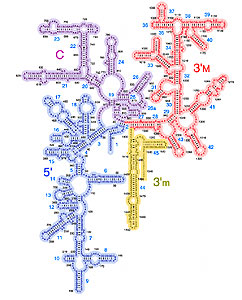
Back home, after your trip to the deep-sea hydrothermal vent, you want to determine what genus of bacteria you have in hand. You can use PCR to amplify the gene for ribosomal RNA (the gene isolated and sequenced by Woese from so many organisms when he constructed his “Tree of Life”). Then, you can choose conserved regions of the rRNA gene for primers. With adequate DNA from PCR, you could sequence the gene and compare it with millions of known rRNA gene sequences using a computer database. (See the Genomics unit.)
Alternately, you might want to ask if a microbe carries out a particular form of metabolism. Given the DNA sequence for a protein involved in a particular metabolic strategy – photosynthesis, for example – you could construct oligos so that the presence of that gene could be detected using PCR.
How does your microbe withstand the high temperatures of its volcanic environment? This has been a question posed by researchers studying extreme thermophiles for some time. Indeed, organisms have been found that tolerate temperatures as high as 110° C. Some archaea produce unusually high concentrations of thermoprotective proteins (heat shock proteins), which are found in all cells. These proteins help refold partially denatured proteins. Other archaea produce unique proteins that help stabilize DNA. You could use PCR to detect the genes for such proteins in your samples.
As the techniques of molecular genetics are applied to extreme environments we will come closer to understanding the wide variety of strategies that organisms use to survive on this planet… and perhaps on others.
7. Microbes and the Carbon Cycle
We have classified microorganisms, including archaea, based on their sources of energy and carbon. The cycling of carbon between carbon dioxide and organic compounds is of considerable ecological importance. In addition to eukaryotes (such as plants and algae), autotrophic bacteria (such as cyanobacteria) play an important role in the fixation of carbon dioxide into organic compounds. Consumers, in turn, use organic compounds and release carbon dioxide. Decomposition of plants and animals and their constituent organic compounds is carried out by a large number of bacteria and fungi.
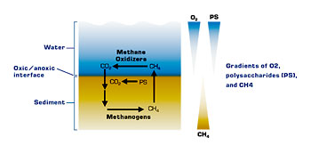
What is taking place in a swamp where you see marsh gas bubbling up though the ooze? A carbon cycle, based on one-carbon compounds, is taking place in the sediments and overlaying water of such freshwater environments. The anoxic sediments harbor archaea, which produce methane as a byproduct of energy metabolism. The methane rises from the sediment

Figure 5. Methanogens are intolerant to oxygen so they thrive in anoxic sediments. The methane they produce is a carbon and an energy source for methane oxidizers in overlaying water.
and moves into the zone above it. This upper area contains enough oxygen to support methane oxidizers, bacteria that use methane as a source of carbon as well as an energy source.
Methane (CH4) is a greenhouse gas and, according to international agreement, its emissions are controlled. Although it is produced by burning fossil fuel, most enters the atmosphere because of microbial action. How can the latter be limited? One strategy is to drain rice paddies more often, limiting the action of methane producers. Another is to add a layer of soil to landfills to encourage methane-oxidizers. Such approaches to reducing this harmful greenhouse gas are being studied.
8. Microbes and the Cycling of Nitrogen
Nitrogen is an important part of proteins and nucleic acids. This vital nutrient is recycled from organic compounds to ammonia, ammonium ions, nitrite, nitrate, and nitrogen gas by a variety of processes, many of which depend on microbes. Different organisms prefer nitrogen in different forms.

Figure 6. Bacteria are key to the cycling of nitrogen in ecosystems. Different species are involved in decomposition and ammonification, nitrification, denitrification, and nitrogen fixation.
The accompanying figure illustrates nitrogen cycling. Note that nitrification (the conversion of ammonium to nitrite and nitrate) in soil is carried out by two genera of bacteria: Nitrosomonas and Nitrobacter. Denitrification – the loss of nitrate from soil to form gaseous nitrogen compounds (N2O, NO, and N2) – is dependent on other kinds of bacteria.
Some prokaryotes are essential to the nitrogen cycle because of their role in nitrogen fixation, the conversion of nitrogen gas to ammonium ions. These ions can then be used to build amino acids. In aquatic environments cyanobacteria are the most significant nitrogen fixers. In soil some nitrogen-fixing bacteria are free-living, such as members of the genus Clostridium; others live in symbiotic relationship with leguminous plants (such as peas and clover). Symbionts, such as Rhizobium, may contribute ten times more nitrogen to soils than free-living bacteria. As we shall see, these symbionts develop intimate relationships with their host plants that require complex communications.
9. Biofilms
We have formed many of our ideas about bacteria by studying pure cultures – homogenous populations growing in broths. In the wild, however, microorganisms live alongside, in, or on other organisms and often produce proteins not apparent in the laboratory. Bacteria communicate chemically with their neighbors and respond to signals they receive. An understanding of communication among bacteria – including those within bacterial communities — is shaping medical treatments, strategies for bacterial control, and providing a new perspective of the interrelationships between species.

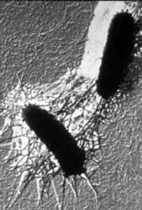
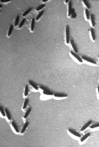
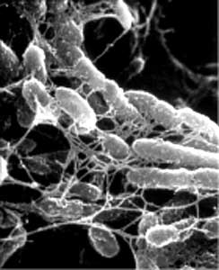
Figure 7. Bacterial cells enmeshed in extracellular matrix material, creating a biofilm
EXTRACELLULAR MATRIX. W.G. Characklis Collection.
One form of bacterial community is the biofilm. An example is the coating of bacteria on your teeth. Biofilms are “living veneers” composed of microcolonies of bacteria, surrounded by a gooey extracellular matrix that the bacteria secrete. A network of water channels provides nutrients and efficiently removes waste products for the bacteria on the surface. Deeper down, cells rely on diffusion for nutrient delivery and waste removal. Oxygen concentrations vary within a biofilm; cells buried deeper can be oxygen-deprived. This variation in environment means that members of a biofilm community, even genetically identical individuals, differ in their metabolic states. In fact, those buried deep within the film are effectively dormant.
“There’s a real transformation that takes place and the bacteria start acting like a community… a whole different organism. And there are significant differences in the level of expression of genes within the biofilm because of the different environments within the microcolonies.”
-Anne Camper, Center for Biofilm Engineering
Biofilms of Pseudomonas aeruginosa in the lungs of cystic fibrosis patients can be life-threatening. The thick mucus that this inherited disorder produces provides a suitable environment for an infection to become established. However, this is not a simple infection. The bacteria organize themselves into a biofilm and, as they do, some become less susceptible to antibiotics. For patients, the result is a prolonged infection that is very difficult to treat.
Why do bacteria in biofilms survive much higher concentrations of antibiotics and disinfectants than free-living organisms? One reason involves the dormant bacteria in the biofilm. Many antibiotics – penicillin, for example — act only on actively growing cells. Cells that were dormant can serve to reestablish a biofilm once the antimicrobial is no longer present.
Another mechanism for survival is the layered nature of a biofilm. The effectiveness of a disinfectant, such as bleach, is depleted as it acts on outer layers of the film; bacteria located in inner layers may survive. A third mechanism for survival involves the generation of proteins that provide antimicrobial resistance, such as enzymes that inactivate hydrogen peroxide. Some biofilms are able to manufacture larger quantities of such enzymes so they become more resistant than planktonic (free-floating) bacteria.
10. Biofilm Formation and Bacterial Communication
How do biofilms form? The formation of a biofilm requires coordinated chemical signaling between cells. Unless an adequate number of neighboring cells are present, the costs of biofilm production to an individual bacterium outweigh the benefits. Thus, a signaling process benefits the bacteria by allowing them to sense the presence of neighboring bacteria and respond to varying conditions. The process by which a bacterium does this is called quorum sensing.
Quorum sensing uses signaling molecules, known as autoinducers. These are continuously produced by bacteria and can readily diffuse through the cell membrane. When elevated numbers of bacteria are present in an area, the concentration of autoinducers in the region will be higher. Autoinducer molecules (which include certain peptides and compounds known as homoserine lactones) can interact with specific repressor or activator sequences in DNA. The presence or absence of the autoinducer thus controls the production of mRNA, and therefore protein. These proteins are encoded by dozens of genes, including the genes for biofilm production. Laboratory strains of P. aeruginosa lacking the gene for a specific homoserine lactone will not develop into normal biofilms but pile up into a disorganized heap.
From the bacteria’s perspective, intracellular signaling has many advantages. Microbes often produce antibiotics that inhibit the growth of competitive species. Intracellular signaling not only brings bacteria together in biofilms, it also regulates the coordinated delivery of high doses of these antibiotics from the denser bacterial population. It also helps bacteria coordinate the release of virulence factors (such as disease-causing toxins) to overcome animal or plant defenses. Signals between bacteria in close proximity, as in a biofilm, also seem to enhance bacterial mating and the acquisition of novel DNA by transformation, both of which increase bacterial diversity.
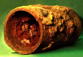
11. Impact of Biofilms on Humans
What is the impact of biofilms on humans? Most are benign, like the slippery coating on a rock in a stream, but others can cause serious problems. For example, biofilms contribute to corrosion in metal piping and can reduce the flow of fluids necessary for many industrial applications, including power generation. A particular concern is the contamination of medical devices such as urinary catheters, hemodialysis equipment, and medical and dental implants. Biofilms that develop on these devices can increase the risk of patient infection. The recognition that biofilm formation contributes to disease extends beyond the Pseudomonas infections suffered by cystic fibrosis patients. Tuberculosis, Legionnaire’s disease, periodontal disease and some infections of the middle ear are just a few examples of diseases that involve the formation of biofilms. The Centers for Disease Control and Prevention estimates that biofilms account for two-thirds of the bacterial infections that physicians encounter.
Several strategies can be used for attacking biofilms. For example, one might interfere with the synthesis of the extracellular matrix that holds the film together. Scientists are investigating coating medical devices with chemicals that hinder matrix formation. Another strategy involves inhibiting the adherence of biofilm cells to their substrate. Identifying chemicals that bind to cell surfaces, stopping the formation biofilms before they begin, is also an ongoing interest of researchers. Targeting the molecules that biofilm bacteria use to communicate is a third tactic.
In 1995 Peter Steinberg of the University of New South Wales, Australia, realized that the fronds of a red algae growing in Botany Bay are rarely covered with biofilms. He determined that the algae produce substituted furanones, chemicals that resemble the acylated homoserine lactones necessary for bacterial communication. Evidently, the furanones bind to bacterial cells, thereby blocking the ability of the cells to receive the signals for quorum sensing. Although these compounds are too toxic for human use, similar compounds are being investigated for inhibiting the Pseudomonas biofilms that form in cystic fibrosis patients.
12. Communication Between Bacteria and Eukaryotes

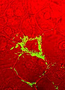
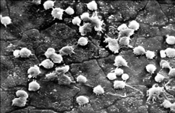
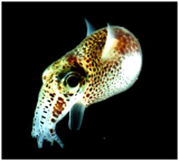
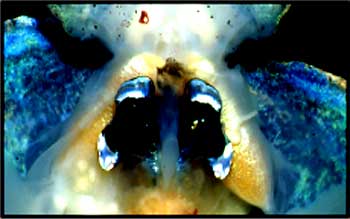
Figure 8a. The luminous bacterium Vibrio fischeri colonizes the light organ of the squid Euprymna scolopes, providing camouflage.
M. McFall-Ngai and E. Ruby, EUPRYMNA SCOLOPES.
Bacteria also communicate with plants and animals. One striking example involves the Rhizobium bacterium, which helps fix nitrogen for legumes (such as pea and clover plants). This bacterium colonizes root hairs in specialized nodules built by the plant. Before the plant and bacteria ever come into contact, they are communicating. The plant sends out chemical signals,
known as flavonoids, which penetrate Rhizobium cells and stimulate a gene-activating protein. The protein then switches on bacterial genes so that other proteins, such as Nod factor, are produced. Nod then stimulates the plant to form nodules.

Figure 8b. The luminous bacterium Vibrio fischeri colonizes the light organ of the squid Euprymna scolopes, providing camouflage.
M. McFall-Ngai and E. Ruby, EUPRYMNA SCOLOPES.
Another example is the signaling between the luminous bacterium Vibrio fischeri and its host, the squid Eupryman scolopes. These bacteria colonize a specialized light organ on the squid, providing camouflage. The squid is a nocturnal forager;
luminescence from the bacteria erases the shadow that would normally be cast from above by the moon’s rays. Quorum sensing molecules allow the bacteria to turn on light production only when the colony has reached adequate density. However, the bacteria do not just communicate with one another — their chemical signals spur maturation of the light organ. Hatchling squid raised in sterile seawater do not develop the pouch that eventually houses the bacteria.
Like the Dr. Doolittle of fiction, who had the remarkable ability to talk with animals, scientists of the future will be continuing studies into the language of microbes.
13. Microbes in Mines
Pyrite (FeS2), otherwise known as “fool’s gold,” may not look like lunch to you but it does to the chemolithotrophic bacteria Acidithiobacillus ferrooxidans(formerly Thiobacillus ferrooxidans). These bacteria extract energy from the oxidation of ferrous ions (Fe2+) to ferric ions (Fe3+). Pyrite is one of the most common forms of iron in nature, and is very common in bituminous coals and in many ore bodies. When pyrite is exposed, as in a mining operation, it reacts with oxygen to generate ferrous ions, sulfate, and hydrogen ions.
2FeS2 + 7O2 + 2H2O —> 2Fe2+ + 4SO42- + 4H+
Ferrous ions – lunch! And, the hydrogen ions generated in this reaction do not faze A. ferrooxidans. This acidophile prefers a pH below 3.5. It is able maintain a relatively neutral internal pH by actively pumping protons between the cytoplasm and external environment against a steep pH gradient.
Acid mine drainage, which causes serious ecological damage to rivers and lakes is in part a result of the presence of A. ferrooxidans. The ferric ions generated by the bacteria are soluble in the acid environment and easily react with additional pyrite.
FeS2 +14Fe3+ + 8H2O —> 15Fe2+ + 2SO42- + 16H+
The additional acid formed from this reaction is just one of the resultant pollutants. The ferric ions (Fe2+) that are generated precipitate in a complex mineral called jarosite [HFe3(SO)4,(OH)6]. The unsightly stains in mine drainages, called “yellow boy” by U.S. miners, are jarosite.
Acid mine drainage and its associated pollution do not form unless pyrite is exposed to oxygen. Only upon mining does the initial reaction generating ferrous ions provide an environment in which A. ferrooxidans will thrive.
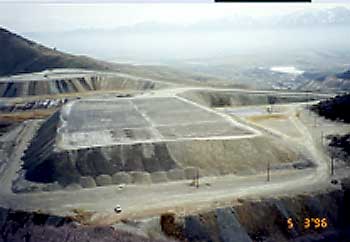
To reduce toxic metal content in acid mine drainage, scientists are turning to sulfate-reducing bacteria, which occur naturally in anoxic soils. These bacteria use sulfate as an electron acceptor instead of oxygen, in a form of metabolism known as anaerobic respiration. Hydrogen sulfide is generated in the process. At a bioremediation site in southeast Idaho Dan Kortansky and his colleagues set up a series of ponds separated by berms (embankments) of crushed limestone, straw, and manure. The goal was to convert the sulfate in the drainage to sulfide. This reacts with the dissolved metals to form metal sulfides, such as ferrous sulfide. The limestone in Kortansky’s bioremediation system lowers the pH as metal-laden water passes through the berms and sulfate-reducing bacteria thrive. Results have been encouraging. Iron concentrations in the drainage at this site were reduced 65% and copper residues were reduced by nearly 100 percent.
14. Microbial Leaching of Ores
Pyrite is not the only mineral oxidized by A. ferrooxidans. Metals such as copper are often present in ores as sulfides. A. ferrooxidans can convert the sulfide chalcolite (Cu2S) to covellite (CuS) to obtain energy. Copper miners take advantage of this metabolic step during the microbial leaching of low-grade ores. Cu2S is insoluble but can be converted by a series of steps (some of which involve the bacteria) to soluble Cu2+ ions. Copper metal (C0) is then recovered when water, rich in copper ions, is passed over metallic iron in a long flume (Fe0 + Cu2+ –> Cu0 + Fe2+).
In heap leaching, a dilute sulfuric acid solution is percolated through crushed low-grade ore that has been stacked on an impervious pad. The liquid coming out of the bottom of the pile, rich in copper ions, is collected and the metal is precipitated by contact with iron (as above). The liquid is then recycled by pumping it back over the pile. Three different oxidation reactions take place within the ore pile:
-
- Cu2S + O2 –> CuS + Cu2+ + H20 is accomplished by bacteria
-
- CuS + O2 –> Cu2+ + SO42-is accomplished by both chemical and biological processes
- CuS + 8Fe3+ + 4H2O –>Cu2+ + 8Fe2+ + SO42- + 8H+is a chemical reaction
The resultant Cu2+ is recovered from the solution when it reacts with iron and the Fe2+, which enters the solution, is oxidized (again by A. ferrooxidans) to Fe3+. After oxidation this solution is delivered once again to the ore heap. The last oxidation, dependent on the bacteria, provides the Fe3+ that drives step 3.
The mining industry has increased biological leaching techniques for various reasons, including environmental concerns related to smelting, the decline in the quality of ore reserves, and difficulties in processing. This new interest has motivated increased research. We now know that ore heaps contain a much wider range of organisms than previously thought. In fact, a succession of microbial populations occurs during the leaching of sulfide minerals. Heterotrophic acidophiles belonging to the genera Acidiphilium and Acidocella are found frequently, often in close association with A. ferrooxidans. These heterotrophic species probably scavenge organic molecules that are metabolic byproducts of the chemolithotrophs. Perhaps this association is detrimental, or perhaps it helps A. ferrooxidans thrive by removing wastes.
Research continues into the composition of bacterial communities that occur naturally in bioleaching activities. Because ore heaps get quite hot during bioleaching, scientists are also asking whether novel bacteria — perhaps thermophiles from Yellowstone or deep-sea vents — might be seeded onto heaps to provide more efficient biomining.
15. Coda
There are about 5,000 known species of prokaryotes, but scientists estimate that true diversity could range between 400,000 and 4 million species. Each has adapted to its particular environment and each performs many roles. Some of these roles are essential to sustaining entire ecosystems. But what is a prokaryotic species? Microbes, which reproduce asexually, cannot be thought of in terms of reproductive isolation. The advent of molecular genetics has brought with it new approaches to defining the concept of species. Some bacteriologists are differentiating prokaryotic species based on their rRNA sequences. If organisms possess rRNA sequences that differ by more than a certain proportion (usually three percent), these bacteriologists consider them different species. As new molecular genetic approaches to the study of microbes are developed, scientists will find additional ways of describing the vast diversity of organisms that make up a parallel, albeit invisible, part of our world.