


Rediscovering Biology: Molecular to Global Perspectives
Neurobiology Expert Interview Transcript: Wolfhard Almers, Ph.D.
 Senior Scientist, Vollum Institute
Wolfhard Almers, Ph.D., is a senior scientist at the Vollum Institute in Portland, Oregon. Neurons release neurotransmitter packed in vesicles in a process called exocytosis. Almers uses evanescent field microscopy to visualize and examine the events and molecules surrounding exocytosis, including vesicle docking, membrane fusion, and recycling.
Interview Transcript
Wolfhard Almers, Ph.D., is a senior scientist at the Vollum Institute in Portland, Oregon. Neurons release neurotransmitter packed in vesicles in a process called exocytosis. Almers uses evanescent field microscopy to visualize and examine the events and molecules surrounding exocytosis, including vesicle docking, membrane fusion and recycling.
What’s the main focus of your research here?
The main focus of my work is to study the mechanism of transmitter release by nerve cells, and how nerve cells clean up after they’ve released their transmitter, basically.
What’s a synaptic vesicle?
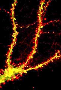
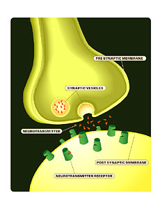
So the way neurons talk, they both listen at the same time and talk at the same time – unlike people, who have a hard time listening and talking at the same time. But neurons can do that. And they talk and listen via structures called synapses. Synapses are places where two neurons come very close to each other. They’re very small: maybe one-hundredth the size of a human hair. And the synapse has two parts. One part belongs to the neuron that’s talking; it’s called the presynapse. The other part belongs to the neuron that’s listening, and it’s called the postsynapse. And the presynapse talks by releasing small molecules called transmitters. There are maybe five or six true neurotransmitters known. In order to release them and do it fast, they prepackage them in vesicles inside the cell, inside the presynapse. So a vesicle is like a soap bubble — very small — and it is surrounded by a membrane, just like a soap bubble is surrounded by a film of soap, and inside it is the neurotransmitter. And the entire presynapse, or presynaptic neuron terminal, is also surrounded by a membrane. And the transmitter is released when the vesicle fuses its membrane with the membrane that surrounds the presynaptic terminal. So that’s called fusion, or exocytosis. You know when you blow soap bubbles, sometimes you blow one, and then there are actually two stuck together that come out? Very rarely, they actually become one, and that is sort of what happens when a synaptic vesicle membrane fuses with the membrane that surrounds a synaptic terminal. And the outcome is that what was inside the vesicle, the transmitter gets to end up outside the cell. That’s called exocytosis, and that’s how neurons release transmitter.
Is this basically the opposite of endocytosis?
So endocytosis is what cells do to clean up after they’ve performed exocytosis. When a vesicle fuses with the plasma membrane, the membrane that’s part of the vesicle gets to be part of the plasma membrane. Sometimes the cell might like to have that, but sometimes, if there’s too much of it, then the cell will run out of vesicles. So this membrane that belongs to the vesicle originally, and is now in the cell membrane, has to be retrieved, and the mechanism for that is called endocytosis, the opposite of exocytosis.
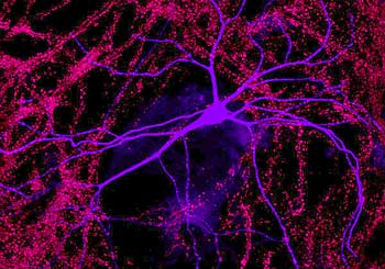
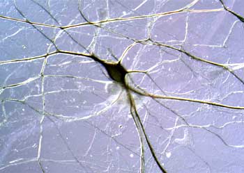
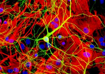
Could you explain the main parts of the neurons and what they do?
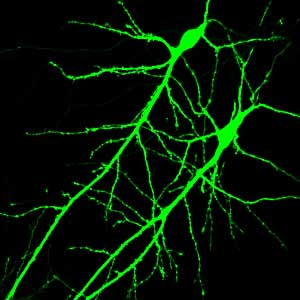
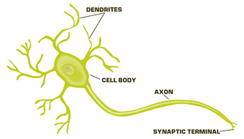
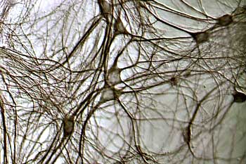
So neurons are cells that have two ends. There are also round cells, like your blood cells. They’re just round. They have no ends at all. But a neuron has basically two ends. The one end, in many neurons, is called a dendrite; basically a tree – very, very small, of course — that branches in many directions and this dendrite is a place to form synapses. So through a dendrite, a neuron can receive maybe anywhere from ten to ten thousand inputs. The other end of the neuron is the axon, which also branches, and the end of the axon touches the dendrite of another neuron. And there can be many branches of these axons; anywhere from one to ten thousand. So neurons can make many connections and will make many connections. And the synapse, where the tip of an axon branch joins a dendrite – it’s about one micron in diameter, about a hundredth the width of a human hair – that synapse is sort of the equivalent of a transmitter, if you want to compare the brain with a computer. The big exception is that, when the hardware for a computer changes, that means it breaks, we throw it away. But synapses change all the time. While we are speaking, and every morning you wake up, you’re the same person-almost. You’re never quite the same person because, through the day’s experience, your synapses will have changed as a result of neuro-transmission.
Could you touch on the role of myelin?
Sometimes neurons want to transmit messages over long distances. The longest neuron in our body goes from our head to our toe. It senses if somebody steps on your toe, then we find out about that through neurons of that kind. And over such long distances, in order for electricity to work, you have to surround the axon, which is a piece of the cell, with an insulator, just like you would surround a cable with an insulator. Neurons use electricity to transmit information, especially over long distances; it’s fast for no other reason. And that’s what myelin is for. If myelin breaks down – there are diseases when it breaks down – then you can’t feel what’s going on in your toe or you can’t move your leg because you can’t move, you can’t contract your muscles.
Give us some idea of how many neurons you would find within the brain.
There are probably a billion neurons in our central nervous system, and about a trillion connections.
Each single neuron makes how many connections?
On average, they make about a thousand connections, very roughly. But there are neurons that send a signal to only one other cell. And there are other neurons that get input from only, you know, maybe ten cells. So it varies quite enormously. There are big neurons and small neurons.
So the neuron is the “centerpiece” of brain function?
It probably is the centerpiece, because there isn’t anything that a neuron does that some other cell cannot do and sometimes better. And what’s special about them is that they’re wired together and in a very complicated set of connections. And we’re only beginning to understand how complicated it is and how it changes.
Give us the process of how a message is sent from one neuron to another.
Neurons, as do some other cells, they release transmitter. They have their vesicles fuse in response to an intracellular signal, which is calcium. So, normally, the calcium concentration inside a cell is very low. Way, way lower than you could taste. And when an axon transmits an electrical impulse, the electrical impulse opens channels in the membrane of the presynapse. Channels are like little holes, which selectively let through ions, such as calcium or sodium; sometimes at the same time, sometimes just one at a time. And the calcium flowing in through those channels raises the calcium concentration in the terminal for a very short time, for a millisecond or so. And the vesicle that sits right next to that channel suddenly sees the high calcium, and that is its signal to fuse with the plasma membrane. And then there is a connection between the inside of the vesicle and the outside of the cell, or of the terminal, and through that connection the transmitter passes to the outside of the cell. And once outside the cell, the transmitter is ‘captured’ by molecules of the postsynapse. And those molecules, called receptors, bind transmitter molecules, and they then know to tell the postsynaptic cell that it has received transmitter. And it does that through the opening of other kinds of ion channels. So the fast aspects of communication happen through ion channels. There are ion channels in the presynapse, where the trigger for release comes into the presynapse and ion channels in the postsynapse that result in the electrical potential changing. And the neuron then looks at that potential and sees what it should tell to the neurons that it talks to.
How did you get into this work?
There’s probably not a single intelligent human in this country who is not interested in how the brain works, because after all, we are taught that the soul resides in our brain, and the person that we are is in our brain. That was not always believed. The Greeks thought the seat of the soul is the intestines; hence, your gut feeling. Others thought it’s the heart, but now we think it’s the brain. Certainly, if the brain is not functioning, it’s very difficult to talk to somebody and communicate with them. So most people are interested in nerve cells and in brains, and so was I. I guess I was interested in how – you know, how we recognize our grandmother. Questions like how a cat knows that this thing there is a mouse and that it’s good to eat. And very soon I became convinced that these questions are too complicated to study. This was 20 years ago. They’re now becoming possible to study. And it was first necessary to understand the little bits that a central nervous system is made of. Just like a computer is made out of transistors, so our interest became how synapses work and how neurons and synapses release transmitter.
Tell us about the research that led you to capture neural communication on video.
So how did we get there? We were always interested in capturing single events of release by one method or another. And the best method for that was really electrical recording. So we, and others, have done electrical recording from single neurons, or cells that behave like neurons. There’s one problem with the electrical recording, and that is that all you can ever find out is when exocytosis or when the fusion event happens. What happens before, or what the vesicle does before it fuses, you can’t tell. Electrical access doesn’t let you do that. But you can tell if you can see vesicles and watch them, and this is what we wanted to do, and we started working on small spherical cells, as big vesicles, and then progressed to working on neurons, which make the smallest vesicles any cell makes. And that’s where the microscopes that we use become useful.
What’s significant about what a vesicle does before its released?
It’s of interest, first of all, to understand the molecular mechanism. Because, as you might imagine, a vesicle swimming up against a plasma membrane, it doesn’t just fuse. It’s very difficult for that to happen. So it has to assemble a whole bunch of macromolecules at that spot. And then it has to become ready, and then it has to wait for the calcium to come in, and then it can fuse. So in order to tell how long it takes for the vesicle to assemble this complex, or what proteins must be assembled there, it’s useful to be able to watch vesicles before and get signals from them before they fuse. The other thing that’s interesting is, it turns out that in a neuron, fusion is useful and maybe even possible only at some sites on the membrane of the terminal. And the vesicle has to find its way there. And how it does that and how it moves there is, at this point, completely unknown. But it’s very important because there are some presynaptic terminals that have maybe a hundred vesicles. They probably turn them over every few seconds or so in a very tightly regulated way. So it’s important to make sure that those vesicles find the right place, and do it quickly, and then it’s also important to recapture that membrane to make new vesicles again; otherwise, they run out of vesicles.
Did you learn anything surprising or unexpected?
I don’t think we found anything that some person hasn’t said at some point would happen. So in a way, none of what we found was a surprise. But saying that it happens and then seeing it are different things. So one feature that was interesting to us had to do with what happens to a vesicle when it fuses with the plasma membrane. So there are some people who think that it just completely becomes flat, flattens itself into the plasma membrane. Like if you take a drop of oil and drop it on a puddle, you can see the oil spreading over the surface of the puddle. And many people believe that that’s what happens with the membrane of the synaptic vesicle. Others believe that the synaptic vesicle stays as a vesicle, and the cavity of the vesicle stays behind, like a ghost, even after the transmitter has gone out, and that its membrane doesn’t really mix with the cell surface, and that it then just closes off again, gets refilled with transmitter, and can be used again. Both of these are postulated to happen, and may happen in different neurons, and in these first recordings of fusion events, we’ve found that in the neuron that we happened to look at, it was more like the oil drop that spreads on a puddle.
Why did you use goldfish retina?
It’s one of the few places where a presynaptic terminal can be prepared in isolation. Because normally, presynaptic terminals are stuck to the postsynaptic side of the synapse; they’re extremely close together. And with the microscope that we had to use, you can’t see the vesicles coming out of the presynapse, which is where you really want to look from. So we needed a presynaptic terminal that you could plop down on the glass slide and then watch through that glass slide. For most terminals, it doesn’t work because the postsynapse is in the way. But the retinal bipolar neuron can be prepared so that the postsynaptic bits can be made to fall off, and you have the presynapse piece alone, and then you can watch it to your heart’s content. And then you can also watch that release of transmitter or fusion, which happens only at specialized sites, and there are always some that happen to fall on the glass slide that you’re watching. That’s why we use goldfish neurons.
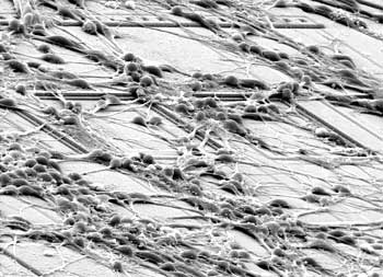
Can you explain what evanescent field and wave microscopy is, and what it enables you to see that you can’t with traditional methods?
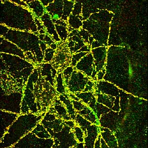
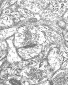
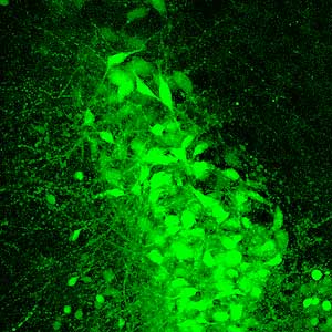
Most small things in biology, most really small things, have been first seen by electron microscopy. You can see single molecules with an electron microscope. And the electron microscope was wonderful. It just shows you exactly what something looks like. And the only problem is that whatever you see is already dead, you know. It’s fixed. It’s been coated with something — a heavy metal and so on, so it’s basically dead. And to see things happening in the live cell, you need to use light microscopy. Light microscopy- the price you pay for that is that it’s harder to see very small things, because the lenses that we use on microscopes, by very deep principles of physics, blur small things so that they look large. A microscope is no real exception to that principle. Now, seeing something small, it’s okay if you can’t recognize its face. If you just see that something is there, that’s already good enough. If you want to tell whether it fuses with the plasma membrane, or whether you want to see it approaching the membrane, or wandering around in other ways. So you don’t need to see its face. You just need to see that something is there. The difficulty in a synapse is that vesicles are packed together very tightly, and if you want to see vesicles, you have to mark them with something that makes them glow, or fluoresce, more precisely. If you look at a synapse in a normal microscope, it may be full of a hundred vesicles or a million vesicles, and all you see is a blur. You know, it’s like watching a bag full of peas and each of those peas glows, and if you watch it from far enough, it will just look like a bag full of glowing stuff. So we had to find a way to look only at a very few vesicles at a time. And one way to do it is to stain only a very small fraction, one percent or so, and then the really critical part is- with this bag full of peas-only to look at the vesicles at the surface of the bag. Then you don’t get confused by the ones behind them. And you can also tell when vesicles approach the surface. And for that, we use an evanescent field microscope.
Explain how this evanescent field microscope allows you to see surface vesicles as opposed to others.
Yes, that is actually not that difficult. In a fluorescence microscope, you see only what you illuminate. Just like at night, you shine your lights at something, and whatever is in that light beam, you see. So with a fluorescence microscope, you illuminate something before you can see it. And the trick is to illuminate only a very thin layer near the surface, and that’s done by a mechanism called total internal reflection. All of us have seen it, basically. So if you drive down a highway on a hot summer day, then the surface of the highway where it’s black and absorbs heat, the air there is hotter. And then it becomes shiny. It looks like you’re looking at a pool of water, and that is the basis of people in the desert seeing water where there isn’t any, because shiny air looks like a pool of water. And what that layer of air does on the highway -it can be very thin; it looks thick, but it’s actually only very thin – is to completely reflect the light of the sun that falls on it. It’s called total reflection. And it happens whenever light goes from a medium that is dense, like glass, and to a medium that is less dense, like water or air. In the case of the highway, the light goes from the cold air, which is always denser, until it hits this hot layer of air on the freeway, so all the light gets reflected. And light consists of photons, so what is reflected is the photons, and that’s what we see. So it turns out that when a photon hits an interface from something dense to something less dense like hot air, photons are a bit like people. You know, they can’t turn around instantly – they will walk for some distance into that rarer medium before they realize that the medium is rare, and then turn around. And because photons are very small, and do things fast and very precisely, that layer is only very thin, only about one-thousandth the width of a human hair. And by illuminating nerve terminals in that way, you can illuminate only the surface and see only the vesicles that are parked there. So with this microscope, the light travels up through the glass slide, which a synaptic terminal is sitting on, and the glass is dense. You know, if you bite on it, it’s hard. And then when it travels through that glass slide and sees the salt solution that is on the glass, or it sees the cell, it notices that that is less dense, and the photons then turn around. And it takes them about a hundred nanometers to turn around. That distance is so small that you don’t get confused by the vesicles behind the ones you’re seeing. So you illuminate only a hundred nanometers deep, which is about two vesicles diameters or so.
What exactly do we see when we see neurotransmission; what’s represented on the monitor?
So what you can see is vesicles that sit there waiting at the membrane, and when the cell is stimulated and calcium goes inside, then these vesicles spread over the cell surface, or the dye in them spreads over the cell surface like the oil drop that you put in a puddle of water. That’s what you can see. And that is a sign that exocytosis has happened. You can also see that not all vesicles fuse when calcium comes in. Vesicles have to have been sitting there for a while, and we can tell now how long that time is. It’s about a third of a second. And there was no measurement of that before. For all we know, it could have been a thousandth of a second or a whole minute. We can see vesicles walking – approaching the plasma membrane-and we are interested in studying the molecules that help it approach. We can test whether it’s actually molecules that are needed to do that, or whether the vesicles can just rattle around, you know. Just the fact that we live in a warm climate and in warm air means that all our molecules are dancing around. Like all the molecules in your body would be dancing around, and still you’re sitting still, and that’s because the molecules don’t dance very far. But if they dance for a hundred nanometers, which is one-thousandth the width of the human hair, you would still be sitting still, but on the distance level that our microscope watches, it’s quite a long distance. So they just could be dancing because they’re warm. But we don’t know.
Could you tell us what the research tells us about the timing of molecular mechanisms?
People have had really wonderful techniques for finding molecules involved in one process and another; so good that they’ve found out thousands of molecules, but they don’t know what they’re involved in. So we are awash in knowledge about molecules, and yet we know less and less about what molecules do and also how they do it and how – even how long it takes them to do it. So information in a living cell about how long it takes for an event to take place is very important. So that’s where the timing is important. What we are interested in now is to actually see the molecules being recruited to where the vesicle sits. So this was kind of a warm-up to finding out about the recruitment, which is something that we’re really much more interested in. We wanted to find which molecules have to be there at the same time to make things happen, then which come first, and which are the stragglers, and so on, and the earlier research sort of put us on the road to doing that.
Are there practical implications for that in terms of a clinical application?
Once you know what roles the molecules play, you can design molecules that go into cells and speed them up or stop them. So that’s the basis of drug therapy in a way or another. And synapses are important for central nervous system disorders because probably in every neurological disorder, something goes wrong with synapses. It’s likely that the synapses are where our information is stored that we remember, and they are where we learn, and also where we forget — that’s just as important. So it’s probably not fair to say that what we do will help patients tomorrow, but as basic research that sets the stage for probably other people — maybe yourselves — to think about what one can do to influence those molecules in a live animal or, ultimately, in humans.
Could you discuss the 1990s as being the “decade of the brain”?
The 1990s were the decade of the brain — I guess that’s stopped now, has it?
I don’t know if that’s necessarily the case.
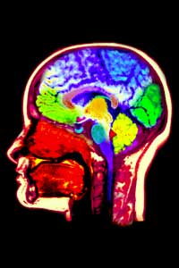
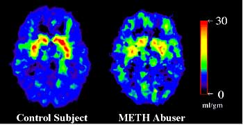
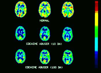
Well, what innovations happened in the 1990s? Probably the biggest innovation, which we haven’t completely taken advantage of, or maybe are just beginning to take advantage of, is to identify by genetic means probably about nearly 50,000 molecules. The human genome encodes about 50,000 molecules. So our cells know how to make 50,000 different molecules. In terms of brain function, probably – I would say the biggest advantage has been imaging of one sort or another. One is using positron emission tomography to image whole brains, and they can do that at a resolution of small bunches of cells. And then of light microscopy, where we can now, in a way, see single vesicles and also see single protein molecules being recruited to vesicles or to other places. And that, in turn, has been made possible by really one advance that sounds trivial, but that is really fundamental; and that is to find proteins that fluoresce. Now, nobody would have thought that proteins could naturally fluoresce. And then one was discovered in a fluorescent screening assay, which is called GFP, or green fluorescent protein. And jellyfish use it to signal to each other–probably. So they have a way to make light, which turned out to be blue. Blue is not propagated well in ocean water, so they make a longer wavelength and they make it green. And the beauty of proteins is that you can find out what the genetic code for them is, and then you can put them into a cell, and then the cell knows how to make that same protein and can actually hook it onto another protein that you might want to watch. And that has been a really, really big advance. And more proteins of that kind are found all the time, so we didn’t really know they existed until about ten years ago, but now people find them in many places. That’s a third big advance for light microscopy that was very important. And many people who have used other methods have now thrown away those methods, and now they all do light microscopy.
Are there other new directions that we may now try to go in?
The new directions that are of most interest to me are ones that we can essentially now see-single molecules or small groups of molecules in action. And that’s important because a cell is small enough. It’s maybe one-tenth the width of a human hair. But a cell, in a way, is like a universe with lots of private corners where things happen, that the rest of the cell doesn’t really find out about immediately, and where things can happen in private. And to watch these events, one needs to look at a small group of molecules in a discrete place, and that is now possible thanks to these fluorescent proteins. The other big advance is that, thanks to advances like microscopy, you can now begin to watch neurons in animals, in living animals. And you can now watch neurons make new synapses, or break old ones down, and test whether it happens at all in an adult animal. So these things are now possible, and I would say within the next five years there’s going to be enormous progress in that.
Do you have any personal impressions of the work that has to do with synaptic plasticity?
We’re seeing things now that we never thought we would see. I guess to me what’s most exciting is to watch things happen in an intact animal because ultimately, whatever you have on a glass slide or in a dish is only in a dish. You never know whether what you see happen in a dish really happens in that way in an organism. It turns out that usually, it’s a pretty good guess. But doing that in an intact animal in the future, I think, seems a wonderful thing to be able to do. And the other thing that we will be able to do is to watch the really elemental things that a cell does, in the living cell. Before, it was possible in a test tube or an electron microscope, and now we can actually watch it happen in the cell. I think we will understand much better why a cell needs 50 proteins to do what seems like something very simple. And you won’t understand that by just listing those proteins, but we will know in the next five, ten years just about everything that a cell does, at that level of detail.
Are there any particular areas that need deeper research and experimentation?
Probably most of what the nervous system does is not understood. And if you had a faint idea as to how the nervous system does what it does, we could build computers that emulate the nervous system, and we’d be ahead. Whoever makes that is going to be ahead financially, militarily, if it has to be. The brain’s processing power is absolutely enormous, and we can’t build computers – we can’t even think about how to build computers that recognize things. So that’s completely unknown. It’s still not clear how we learn what we learn. How we forget is also not clear. Is it genes that get silenced or turned on? Is it molecules that get altered? Is it a mixture of all of these? Is it new synapses that grow? Is it synapses being taken away? These things remain unexplored.
Why we sleep is completely unknown. Everybody knows we need sleep. Why we do it is unknown. Nobody knows whether flies sleep. I bet they do. Why we become addicted to substances is unknown. Why things that we like ultimately deprive us of our judgment if you get too addicted to them. And then, of course, the final mystery, which may never be known, is whether, once you understand what the brain does, do you understand consciousness? How those two will fit together may never be known, because one is philosophical and the other is just looking at things carefully.
Are there any ideas that you want to get across to an audience of high school biology teachers or the general public? I guess the first thing that I want people to know is that doing research is really very exciting. And it’s wonderful, but it’s also sad that half of the people that do research in this country come here from abroad, as I did. So it’s wonderful, in that all these people come here to do research. But in a way, it’s sad that there are too few people who get interested in research in this country. So I guess one thing that would be wonderful to get across is that it’s a wonderful career. It’s very exciting. It is addictive, so maybe – maybe in that sense, it’s not very good. But it’s a very exciting thing to do. The other thing that is very important to me is to understand what drugs do and what they don’t do. And to understand enough about them to tell when somebody’s pulling the wool over your eyes. It’s good to know, for instance, that if there’s a new medicine that’s advertised on TV, that this medicine doesn’t really do anything that some other medicine doesn’t also do, which costs one-tenth of the cost and that is maybe not even as harmful. So it’s good for people to be able to be critical and to understand that, the purple pill, you know, what does it do that its predecessor didn’t also do?
So you think it’s important for people to educate themselves better about treatment and drug options.
The best way to educate yourself is to go on the web. You just go to Google.com and type in whatever drug you’re interested in. The purple pill. Maalox. Ginseng tea. And you will have a whole forum of people who have opinions about it, some better and some nonsense. But ultimately, through that, you can find your way to literature that goes as much into depth as you like. There’s another web site that you can go to, and that is called PubMed. It’s the website of the National Library of Medicine. And there, too, you can type in any subject that you like, and you’ll find the newest references in the academic literature, and it’s all free. And anybody with an Internet connection can do it. You can find reviews that tell you about something in a general way. You can find the latest papers that look at fine details. All of that’s available at this point on the web.
What questions drive your research? What keeps you awake at night?
I try not to be kept awake at night. I guess I would be happy if I could find out how fusion works on a molecular scale, which is really largely unknown for one really simple reason; and that is that we’re really good at finding out what proteins do. The last 20, 30 years were all about proteins. We know their genetic codes. We know how to change them. We know how to tell cells to make them and to make changes in them. We know an enormous amount about proteins, but there are other important molecules that are structurally important, and those are lipids. Membranes are actually made from lipids, and only in part from proteins. And fusion, because membranes fuse, will involve lipids. Our methods to explore those are not really much better than they were 20 years ago, and we need a big advance. I think light microscopy at the single-molecule level or at the level of a small group of molecules will provide that advance, and I guess that’s what I’m after. If I can answer that question I will be very happy.