

Rediscovering Biology: Molecular to Global Perspectives
Microbial Diversity Expert Interview Transcripts: Anne K. Camper, Ph.D.
 Associate Professor of Civil Engineering; Associate Dean for Research and Graduate Stud
Camper is an associate professor of Civil Engineering and the associate dean for Research and Graduate Studies at the College of Engineering at Montana State University in Bozeman. Her research interests include bacterial attachment to surfaces, and biological treatment of drinking water and microbial regrowth in drinking water distribution systems.
Interview Transcript
Anne Camper, Ph.D., is an associate professor of Civil Engineering and the associate dean for Research and Graduate Studies at the College of Engineering at Montana State University in Bozeman. Her research interests include bacterial attachment to surfaces, and biological treatment of drinking water and microbial regrowth in drinking water distribution systems.
What’s new in the study of bacteria?
Over the past hundred years or so we’ve always thought of bacteria as being independent free-floating single organisms. Recent advances have shown that probably the predominant life form for bacteria is in biofilms, either as bacteria attached to the surfaces or attached to each other. I think we’ve changed our viewpoint considerably in the last ten years.
Do the majority of bacteria live in biofilms?
That is starting to be the belief right now. Obviously, there are certain environments where free-floating suspended organisms predominate, but in many cases in nature we find them attached to surfaces.
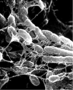
What is a biofilm?
A biofilm is a consortium of organisms attached to a surface usually at an interface and that means that an air-water interface and air-oil interface or sometimes a water-air interface. Usually they’re attached to some sort of hard surface although it could be tissue in the human body as well.
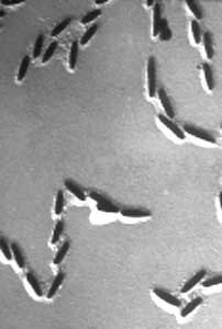
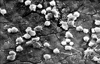
Is the first step in biofilm formation to find a place to attach?
There are two concepts on initial attachment. One can be an almost chemotaxis approach where the organisms sense that there’s an accumulation of nutrients on the surface and they’ll actually begin a random walk in that direction. They are attracted to that surface because they recognize something on that surface that may serve as a nutrient source for them.
The other idea is that they just physically bump into the surface by the action of the local hydrodynamics. So they’re transported to the surface through the fluid flow so there could be what’s called a “specific” or a “nonspecific attachment” once they reach that surface based upon the interaction of the cells with the surface.
Do the bacteria have a sense of touch?
I don’t know that they have a sense of touch, but they do seem to be able to locate a surface and know that they’re in association with it, specifically when we’re talking about specific attachment because there are receptors on the cell and some molecules on the surface where the organisms will actually make an interlocking mechanism between the cell and that surface and then remain in contact with it.
Do they roll around to find out whether they want to stay?
It depends upon the environment in which they find themselves but what we’ve noticed with most motile bacteria-those are the ones that have flagella-is once they come in contact with the surface, they don’t make an immediate permanent association with that surface. They may move around on that surface for a period of time and once a certain time period has passed they’ll make an irreversible “decision” to remain on that surface. At that point in time it appears that some of the physiological responses of those organisms changed and they now become a biofilm cell, as opposed to a suspended cell.
What is the change that happens to make a suspended cell turn into a biofilm cell?
It appears that there are specific genes that are up-regulated or certain genes that maybe down-regulated when the cell comes in contact with the surface and a lot of those genes are going to be different depending upon the individual species. But in the case of P. aeruginosa, for example, those organisms lose their flagella and they start producing pilli instead. It also appears that some of their metabolic functions change. They also start to produce the extracellular polymeric substances that act as that glue that stick them to the surface at that point.
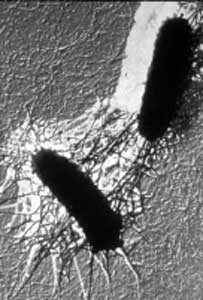
What is the difference between flagella and pilli?
A flagellum is a structure that allows the cell locomotion, usually in a suspended culture. So they will use those for chemotaxis, to move from one point to another.
The pilli appear to be more related to cell interactions and cell surface interactions.
They allow the cells to remain in contact with the surface and also to form communication structures perhaps between each other.
Once the cells stick, what do they do?
Once the cells have decided to remain on the surface there does appear to be a fairly substantial change in gene expression and that gene expression does include things like expression of the genes responsible for the polymers that stick the cells to the surface and a variety of other functions, some of which we do not clearly understand at this point in time.
What are the polymers?
The EPS or Extra Cellular Polymeric Substances is the glue that holds the cells to the surfaces. In some cases, we understand what that EPS material is. For example, P. aeruginosa produces alginate. It may produce other compounds as well. In other bacteria we really do not have that EPS clearly defined but it’s typically composed of carbohydrates, although there may be some proteins and there’s even some evidence now that DNA may be involved.
Does the biofilm use the EPS as a source of nutrients?
The EPS could potentially act as a reserve substance for nutrients. We do know that it helps trap substances from the environment. It’s one of the advantages that a biofilm cell would have. This EPS can act as an attractant or it can actually hold nutrients from the environment allowing the cells to have some reserve if hard times come later on.
How do the bacteria know to congregate?
It appears that some of the initial stages in biofilm formation are not governed by cell signaling. They’re governed more by the environment and then either programmed or chance encounters with the surface.
Once they’re there and there’s a sufficient accumulation of certain molecules like cell signaling compounds, then we start seeing some additional changes in physiology related to the accumulation of those compounds. Those signal molecules can either affect processes that would enhance biofilm formation or tell the cells that they need to get up and leave the biofilm and become suspended again.
What is quorum sensing?
Quorum sensing is most easily defined as being able to determine the number of cells in the environment or being able to sense the cells in your environment, and that occurs by the excretion of compounds that can be taken up by surrounding cells.
The cell signaling or quorum sensing systems appear to take over when there’s a critical mass of cells that are producing a critical mass of these compounds. So you need to have enough compound produced that it accumulates in the environment to a level that allows for diffusion into the cells, and once that occurs then you can have this transformation in gene regulation.
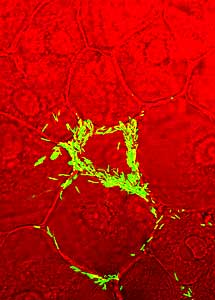
What does the biofilm structure look like?
Biofilm structure is going to be highly dependent upon the environment and the types of organisms. What we usually find, though, is something that’s a little bit complex. It’s not typically a flat layer of bacteria. That used to be the old concept of biofilms that we had. But when you actually look at them you can see towers or pillars, you can see mushroom-shaped structures, you can see porous structures through the biofilms so it actually gives it a three-dimensional confirmation.
What type of tool do you use to see the structure?
There have been two tools that have been incredibly important in our understanding of biofilms. One is the confocal scanning laser microscope. With that we can obtain nondestructive, three-dimensional images and then be able to go back and analyze those images to get information about how the biofilms are put together. So that’s been a very, very important tool.
For some of the work that I’ve been doing proteomics and the ability now to do 2D gel electrophoresis greatly improved our understanding of what’s happening with the individual bacteria in a biofilm system.
Confocal microscopy works to see biofilm structure?
The confocal has all of the limitations of a traditional light microscope. You’re still using a light microscope to collect the information, but instead of using a white light source you’re actually using a laser as the light source and there are different types of lasers that can be incorporated.
With those lasers, you scan down through the biofilms so the excitation on the biofilm is only in a small point in time and space and then you can collect all that information and put it together. What the confocal allows you to do, though, is to get rid of any background scatter so it focuses the light or gets the signal back only from a very small area so you get a very clear image at a single focal plane within a system.
How tall do the biofilm structures get to be?
The structures are highly variable, again, depending on the type of the organism and the type of environment that they’re growing in, but these biofilms can be hundreds of micrometers thick, and those towers could be all the way from the base of the biofilm up to the surface. So it’s highly variable but they can be quite substantial.
Why do some bacteria break away and again form flagella?
What tends to happen later on in biofilm development is that areas within that biofilm become nutrient depleted or there’s waste product accumulation or something else that makes it less conducive for those organisms to stay around in that particular biofilm.
At that point in time, it now appears that there is a re-regulation or an up-regulation of the genes associated with the free-swimming or suspended lifestyle. And what we’ve noticed in P. aeruginosa is that it appears those organisms now start to express the flagella again and down-regulate the pilli and will actually evacuate certain portions of the biofilm and swim back off into the nutrient environment.
Where do they go then?
We assume that they’re going downstream looking for a new place to colonize.
Can you talk about your research?
We’re looking for mechanisms to control the attachment of these organisms to the surface. If we can understand what genes are up- or down-regulated when an organism is in the biofilm mode of growth as opposed to the suspended mode of growth then we may be able to manipulate their attachment or their detachment from that surface.
The stage of development that we’re targeting is going to depend upon the system that we’re interested in controlling. In some instances, we may want to make sure that they don’t attach or at least don’t attach very well. In other instances, if there’s an established infection, we may want to make sure that we can actually cause them to be released but only in the appropriate manner. In some instances release of those organisms in the human body may actually cause problems as well. Such as if you have an organism that is immediately released from a surface and then goes into the bloodstream. That may not be advantageous.
Where are you at in this research?
What we’ll do is grow up these biofilms and it really doesn’t matter what organism we’re working with. It is important, however, at the stage that we’re at right now with the technology that the genome for the organism be already sequenced. We need that to be able to identify the proteins.
Then we’ll go through and collect those proteins and do the 2D gel electrophoresis or the proteomics, identify those proteins that are either up- or down-regulated during biofilm accumulation, and identify those proteins by several different methods that are out there. Then you can go back and determine the genes in the cell that are responsible for the production of those proteins. Once you have that information now you can have an idea of the targets that you need to go for in those individual bacteria.
How do you hope to develop a target to those proteins?
If you can understand the regulatory system for those proteins, you may be able to come up with some mechanisms for interfering with the up- or down-regulation of those genes. You may be also able to develop some vaccines for some specific target proteins that are produced and there are a variety of other potential applications that are out there.
Is this new research?
It is relatively new research. Proteomics is not a terribly old science in and of itself. Of course you’re limited at this point in time to having cells that have been sequenced to get the full advantage and that’s not typical for most organisms yet. We also have had some significant improvements in the technology over the past few years that have made 2D gel electrophoresis much, much easier to perform.
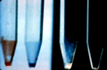
What is 2D gel electrophoresis?
2D gel electrophoresis is the separation of proteins both on the basis of pH and molecular weight, so separation in two dimensions.
Do you have to have that information to combat biofilms?
I think that’s certainly a component of it. There may be some instances where some technologies that we’ve developed against suspended cells will still be effective against biofilms. But to be able to control them in the environments that we’re interested in having that control, we need a whole lot more information on how these cells are different from the suspended organisms. That way we can use more efficient tools or more efficient targets to control those organisms.
How different are free-floating cells from biofilm cells?
What we’ve seen in some of the research we’ve done with P. aeruginosa is a substantial difference in protein expression, either in the number of proteins expressed or the level of protein that’s produced. One of the research projects that we’ve done has demonstrated that a P. aeruginosa grown in suspended culture compared to that grown in a biofilm is as different as two different species grown in suspension. So there appears to be some substantial differences between the organisms.
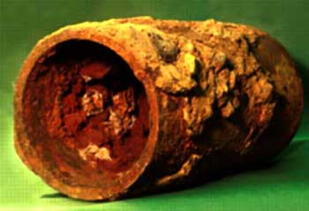
What are the ramifications of this research?
What we’re ultimately hoping for is more efficient mechanisms for controlling biofilm infections. We know right now that using traditional antibiotic therapy can be very ineffective. It’s certainly not giving us the control that we need or that we want. In some instances, antimicrobial therapy is woefully inadequate and I’m thinking of things like cystic fibrosis or if you have an infected prosthetic device in the human body.
In most instances, in the case of the prosthetic device it has to be removed once the infection has been established because traditional treatment methods just don’t seem to work. There are two approaches now that seem to be leading the way. One is designing surfaces for implantation in the body that are less prone to colonization and the other is trying to find mechanisms for making sure that once the infection is there that can be adequately treated.
Is there a difference in gene expression within a biofilm?
There are differences in gene expression in even a single species depending upon where they’re found in the biofilm. An example would be on the internal portion of a large mushroom structure. At that point there will probably be less nutrients and less carbon sources for the cells to grow, probably less dissolved oxygen if it’s an aerobic organism. There may be a greater accumulation of things like cell signaling compounds because diffusion is limited there and there may also be the accumulation of waste products and that’s why individual locations within a biofilm you could have a different expression of proteins or gene regulation that is reflected by the environment at that spot.
What is cell-cell signaling?
I guess the best way to define what would be that these cell-signaling compounds are just small chemical signal entities. They’re out there as individual entities and you need enough of them out there to actually cause the cell to respond so there’s a critical concentration that’s required on the exterior of the cell to make sure that the compounds actually move to the inside of the cell. Once they’re on the inside of the cell, that’s where you see the transformation in gene expression.
How is cell-signalling important to biofilms?
My personal belief is that cell signaling is probably not important to the initial attachment events. Those cells come in contact for a variety of reasons, but once there’s a certain critical mass of cells they may be important. Now most of the information that we have is on single species biofilms. We really have very limited information available on how cell signaling molecules work in mixed-species populations.
Most biofilms are mixed species?
In the environment, most biofilms are mixed species. In a restrictive environment like the human body, we may be looking more limited biofilm populations and it may, in fact, be in some cases single species.
What are some unanswered questions in this field of study?
I think some of the big unanswered questions are still what causes these regulatory changes, what are the key molecules or key systems responsible for this major change in phenotype? I think that’s an area of some very exciting future research that could take place.
Ultimately, of course, we’re interested in being able to manipulate biofilm formation. In some instances, we want to have biofilms form. Bioremediation may be a case in point there. In other instances, they’re not needed, not wanted, and we want to make sure that we can eliminate them so biofilm control and manipulation is the ultimate goal.