


Physics for the 21st Century
Biophysics Interview with Featured Scientist Harald Paganetti
Interviewer: Can you please talk about the history of proton therapy at MGH?
HARALD: The idea came out of the Harvard Cyclotron Lab by Robert Wilson in the late ’40s where he had this idea that particles could be used for cancer treatment. The first patient at the Harvard Cyclotron Lab was treated in the early ’60s—I believe ’62. And then the Harvard Cyclotron Lab was no longer interested in doing basic physics experiments because the machine was getting too old. The machine was built in 1948 and basically the energy region that this machine could cover was no longer of any interest for basic nuclear or atomic physics.
So, they had this idea, together with the Mass. General Hospital, to build a program to use protons for cancer therapy. And that’s how it all started. So, without proton therapy, the Harvard Cyclotron Lab would probably have been shut down in the ’60s. Patients were treated there until 2001 and then we moved to our new hospital-based facility in 2001 and this is really a new facility. So we did not move the Harvard Cyclotron Lab to Mass. General Hospital. We moved part of it but the main part is really new. So, it’s a commercial built facility not an in-house product like the Harvard Cyclotron Lab was.
This is how it worked in the past with proton therapy—it was tried in basic physics labs and patients had to go to labs that were sometimes outside of the city. But in the last 20, 30 years more and more hospitals are getting interested in this treatment option. And now it’s more in the hospital based environment, which has all kinds of advantages because the doctors are going to be around. At the Harvard Cyclotron Lab doctors had to travel there which was not too far in terms of being in Boston. But really in the last 20 years I’d say we have seen this transition from research labs into the clinic.
Interviewer: Quite a few of the staff moved to MGH didn’t they?
HARALD: Yes, most of the staff, actually, moved over from the Harvard Cyclotron Lab to this facility, but there are also a lot of new staff because we have now higher capacity for treating patients.
How many treatments or patients can you treat in a week?
HARALD: We treat about 50 patients a day. This does not translate into 250 patients being fully treated per week because the patient gets on average maybe 30 fractions. So, he needs to come in 30 times because we don’t give the dose all at once because it’s simply better for the healthy tissue which will also receive some dose to get the total dose of 30 fractions accumulating the total dose.
We have two treatment rooms with a gantry. Gantry, meaning that we can rotate the beam around the patient. And then we have one treatment room of two horizontal beam lines where the beam line is basically coming out of a wall and then we either have to move the patient or we are limited in the angles that we can treat the patient with. So, at the old Harvard Cyclotron Lab we could only treat from one angle. And then you have the option, of course, to move the patient around but it’s easier to move the beam around. And that’s what we do with a gantry.
Interviewer: What are you researching?
HARALD: The big advantages of using protons is that your therapeutic ratio increases, which means the dose to the tumor versus the dose to the surrounding healthy structure is actually bigger—the ratio is bigger in proton therapy. In other words, the total dose that you’re delivering to the patient is going to be lower. So, you can use this to your advantage in two ways; one, you can give more dose to the tumor. Let’s say in photon therapy, you might be limited how much dose you can give to the tumor because you have to take into account also that the healthy structure gets some dose. Of course, the ideal case would be you get only dose in the tumor and no dose outside which is unreasonable. You can’t do that because your beam has to reach the target, which is somewhere in the middle of the patient let’s say.
So, the advantage of protons is that you can give less dose to the healthy tissue—or you can maintain the dose to the healthy tissue but at the same time boost the dose to the tumor. So, the integral dose—what we call the total dose to the patient—is lower with proton therapy by a factor of two to three. If we talk about the side effects of radiation—which, of course, occur—the most severe side effect is that patients actually have a small risk of having a second tumor based on the radiation that the healthy tissue received.
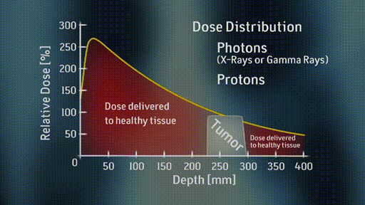
For proton therapy, that risk in theory should be reduced because we’re given a factor of two to three times less dose to the entire body compared to photon therapy. So, that’s why people are interested in using protons basically. The reason why this is happening is a physics reason. It has nothing to do with biology; it’s really physics. If you shoot a photon beam through a patient, it will go into the patient on one side and exit the patient on the other side. So, you have to shoot through the entire patient. And by coming from different angles, you can have your tumor in the focus so to speak, so you get higher doses in the tumor and lower doses outside.
With protons, it’s different. Protons stop in tissue. So, they’re stopped somewhere. So, you can make them by adjusting the energy. You can make them stop in the target. So, you will have dose deposited in the path going through the target, but most of the dose is really going to be in the target. And that’s a big advantage. And so, that’s why people are more and more interested in using protons. Of course, this improved accuracy has not only got advantages. Otherwise, we wouldn’t have to do research. The disadvantages, if you’re more precise, you’re actually more prone to any uncertainties or error.
For example, in photon therapy, if you were to misalign your patient by 3 or 4 or 5 mm it may not be such a big effect in the photon therapy. So, you may be given 10 percent less or 10 percent more dose, which might or might not be acceptable. But in proton therapy because of the precision, you can actually miss by 50, 60, 70 percent because you’re changing the depth of your dose distribution. And so, this uncertainty is a good thing if you know actually where your dose distribution is. And it can be bad because it may increase your uncertainties in delivering the dose. So, our research tries to improve our knowledge about where the dose is actually deposited in the patient. Obviously, we cannot measure that; or at least it’s very difficult to put detectors in a patient to measure the dose that is being delivered. And even if it would measure it, it would already be too late in a way.
What we’re trying to do is to predict as accurate as possible how the dose distribution that we have planned for a particular patient looks like in the patient. And the way to do that is to use Monte Carlo simulations. Monte Carlo simulations are coming from atomic bomb research that was done in World War II and they used the nickname Monte Carlo for these type of simulations. They were being used in atomic and nuclear physics for years. And then, I believe in the ’60s actually, people in radiation therapy realized that this is a great tool also to be used to predict dose in a patient.
So, the difference from a Monte Carlo dose calculation to an analytical dose calculation is Monte Carlo is more accurate. But it also takes more time. So, routinely when we treat patients, we, of course, have to plan the treatment. Beforehand, we have to find out the correct angles to treat this patient—the correct dose. We have to look into constraints of healthy tissue—how much dose can we give to this organ—not to risk any severe side effects and things like this. So, these predictions are basically based on analytical dose calculation methods because they’re very fast, and they’re reasonably accurate. Reasonably accurate means that if we want to reduce our safety margins, we probably have to find those calculations that are more accurate.
If we treat a tumor, we don’t really treat right to the edge of the tumor. We always add some margin,like a safety margin, because there could be some error in all those calculations. There could be some misalignment of the patient. So, that’s the safety margin that you use around the tumor. So, if you want to reduce that safety margin, which is desirable, because then you treat less healthy tissue—we need a more accurate dose calculation. So, Monte Carlo is that tool to get more accurate information on how the dose is deposited in the patient.
Interviewer: What’s the difference between using protons and using photons?
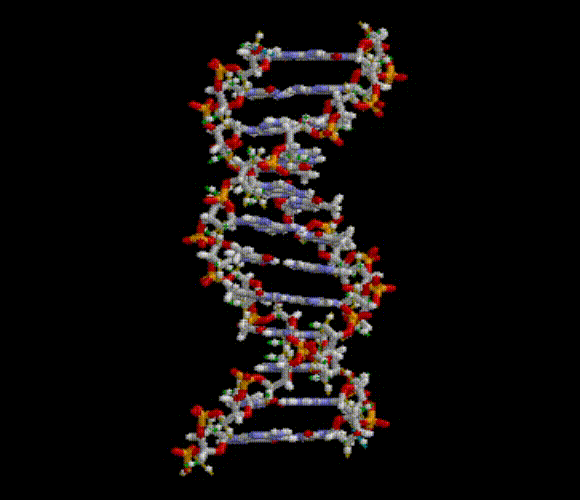
HARALD: What you want to do is you want to destroy the tumor cells. You don’t want to leave behind a single tumor cell. Your dose needs to be actually quite high. Well, for the organs at risk, of course, it’s a different story. Here you’re more concerned about mutation that could lead to severe side effects because the dose that you’re giving to the surrounding tissue is usually in an area where you’re not so much concerned about killing cells, but more concerned about severe side effects. So, you can have interacting with the DNA that will destroy cells and things like this. That’s really what you’re trying to achieve—to dump as much energy into this tumor that the cells are being killed.
Well, the difference between photon radiation and proton radiation is really physics. I mean it’s in part biology, but let’s talk about the physics first. Photons are uncharged so they deposit a dose by a secondary electron, which have a negative charge. And protons are positively charged. And from that difference alone—there’s also a difference in mass, of course—you can guess that the physics interactions of protons are very different than the physics interactions of say photons or electron beams. That’s one of the reasons why dose distribution in the patient looks so different when we treat with protons or even heavy ions, which are also being used for cancer treatment compared to photons and electrons. And that is one of the reasons why protons have what we call depth dose distribution—with dose as a function of depth in tissue—which ends up in the Bragg Peak.
So, most of the energy is actually deposited when the proton slows down; whereas in photon treatment, you get most of the dose at the entrance to the patient. And then it decreases almost exponentially.
If a particle hits a cell, what basically happens is the same as if a particle hits any other material. So, you’re doing ionizations. You’re kicking out electrons—and these electrons deposit energy in tissue—and it is this energy being deposited that can, for example, damage the DNA. And that’s how you get radiation effects either of mutation or even cell kill.
Interviewer: What are the dose differences between proton and photon therapies?
HARALD: One of the differences between protons and let’s say photons is there’s a slight difference in the biological effectiveness. Eventually, we’re not interested really in physics; we’re interested in biology. And we just know from our clinical experience that we need a particular dose to kill a cell, or to kill a certain tumor, or to treat a patient. All of our prescription doses are based on clinical experience; they’re really not based on theory. So, what we know is that protons are about ten percent more effective than photons.
To give an example: if you would treat a patient with 70 gray on the photon side, and you would transfer that patient to a proton treatment, that patient would receive only 63 gray of protons because we know protons are ten percent more effective. But the ten percent is really only an average value. There are some variations—and we know that. But these variations are not taken into account when we treat these patients because there’s still some research being done on how big these variations are. We believe that they are probably in the order of five to ten percent, which is actually quite high because we try to treat these patients with dose to within two percent accuracy. But there’s still research being done on the experimental side but also on a theoretical side how to model these biological effects so that we can predict more accurately what the biological effective differences are between different radiations.
With protons across the board ten percent is a good average value. But we’re trying to be more accurate to reduce the safety margins. The more accurate we know the biology, the more accurate we know these variations, the more we can spare healthy tissue because we can reduce those safety margins that come out of these uncertainties.
Interviewer: What experiments are you conducting now?
HARALD: We’re doing a lot of research in proton therapy to change the way we deliver the beam. So, right now, when the beam enters the treatment room, it’s a very, very small beam, which we call a pencil beam because it’s very fine. It has about a diameter of let’s say a centimeter, something like this. In order to cover the tumor which is bigger than that usually, we have to scatter the beam to make it a broad beam, and we also have to use apertures and compensators. And we also have to add up different Bragg Peaks to make it a flat dose distribution across the tumor. So, there’s a lot of hardware involved in shaping these fields. And the disadvantage of a lot of hardware is that you get a lot of scattered radiation.
So, the treatment being delivered, which is really not that new—people have looked into this for years now, is called beam scanning. What we do is actually use that fine pencil beam that enters the treatment room directly. You have two scanning magnets in the treatment head that can scan this fine pencil beam in an X and Y direction. So, it’s like on your old TV screen where you would scan an image by going through the different levels and you basically scan the particular depth of your tumor and then you would use the energy and you would scan the next layer, you would use the energy, you would scan the next layer and so forth. So, in that way, you could fill this tumor with small little dose spots and cover your dose distribution.
Now, this has obviously an advantage because you have less hardware in the treatment head, but it offers an additional advantage in that you’re now being more flexible in how to deliver this beam because in passive scattering, the older technique, you deliver the whole dose distribution at once. Now you’ll basically assemble this dose distribution by using many, many different spots, and, of course, you have a control that not only can you deliver an inhomogeneous dose distribution if you want to, but you also can shape dose distribution in a more complex way. So, you’re basically gaining one additional degree of freedom. That’s called intensity modulated proton therapy, which enables us to treat from a different direction—as we do with the passive scattering technique. But now from each direction, we can administer an inhomogeneous dose distribution. The total dose adds up to a homogenous dose distribution. But by having these single fields differ in the dose distribution, we gain our ability to stay away from critical structures. So, the main reason for doing beam scanning is really to give us one more degree of freedom to save critical structures or to give less dose to those that are at risk.
Interviewer: Why are magnets helpful in proton manipulation?
HARALD: The advantage of protons is that they’re positively charged, so you can use magnets to move them around so to speak. What we’re doing with pencil beam scanning, we have a small pencil beam of protons going into the treatment head, and then we can deflect the pencil beam with a magnetic field, and we can basically have that pencil beam hit a particular small area of the patient very precisely. By changing the energy of the protons, we can also precisely predict how deep the beam goes into tissues. So, this gives us three degrees of freedom. We can deliver a high dose burst to any region in the human body, which ideally would be in the tumor. So, with our scanning magnets, we can really pinpoint our little pencil beam right to the area where we want to treat the patient.
Interviewer: What is “Monte Carlo”?
HARALD: When you plan a treatment for a patient you have to calculate the dose, and you have to find the optimal orientation of your beam angles and the dose that each beam delivers to the patient. That is called treatment plan optimization. And so, it’s basically mathematics applied to treatment planning.
The dose calculation is being done with analytical methods usually. So most treatment planning programs that you can buy off the shelf is analytical dose calculation. And analytical means that there are some mathematical equations in there that incorporate physics that tell you how the dose is being delivered or being deposited in the patient.
Now, Monte Carlo dose calculation is more accurate simply because it really kind of mimics what’s happening in reality. So, when we do Monte Carlo, we take particle by particle—in our case, proton by proton—and we really track and simulate what this proton is doing while it’s going through tissue. We take a particular proton we enter the patient, we do a step of let’s say a millimeter, and then we look at the physics loss and calculate the probability that something specific happens to that proton, let’s say it deposits a particular dose. Then we take another 1 millimeter step and we do the same thing. We calculate the probability that this proton does something. And it may create a secondary particle. It may create a photon or an electron, for example, and then we also track this particle and simulate exactly what this particle would do in a tissue. So, basically, you’re not covering the dose—the position in one analytical equation—but you’re really doing what’s happening in reality—you’re tracking particle by particle. This is called Monte Carlo simulation. And this is believed to be more accurate and proven to be more accurate than analytical dose calculation methods.
Interviewer: Are Monte Carlo simulations more accurate than analytical calculations?
HARALD: In a Monte Carlo simulation we want to find out whether we need this more accurate dose calculation because it might well be that our analytical methods are accurate enough. And what we’re finding in our research, or what we’re trying to find out in our research is, which areas or which tumors or which sites we need Monte Carlo. And so, what we’re seeing right now is, for example, that if you have interfaces, which means you have bone, you have soft tissue, you have air cavities, the typical scenario for a head and neck tumor, for example, we see differences between Monte Carlo and an analytical dose calculation.
So, that makes us believe that the future is in Monte Carlo and we should continue our research to make it faster, maybe even more accurate, to get it really into the clinic. There might be other areas in the human body when we treat soft tissue tumors where maybe analytical methods are accurate enough. Of course, you always want to be as accurate as possible, but we have to compromise here with the time it takes to do these Monte Carlo simulations at least today.
When I say that Monte Carlo simulations are more accurate than analytical dose calculation, you don’t have to be worried that since we’re using analytical dose calculation, that there’s any danger to the patient at this point. And the reason is that our treatment planners are fully aware of these uncertainties, so they incorporate them in their treatment planning decisions. So, for example, if they know that the dose calculation cannot predict the range to better than let’s say 2 millimeters, they would stay away from critical structures of a 2-millimeter safety margin. Of course, Monte Carlo might eventually help to reduce those safety margins.
Interviewer: What is the future of your research?
HARALD: I think one of the big issues right now is whether we can do personalized treatment planning. Right now if a patient comes in with a particular tumor we would probably prescribe the same dose for that particular tumor if it’s the same tumor type in a different patient, depending on whether we can meet the dose constraints to the organs at risk. But different patients have different genetic profiles so they need more or less dose to cure their particular tumor. Research is currently going into personalized treatment planning, so that we can adopt the knowledge,the genetic knowledge, that we have about patients in the treatment plan in prescribing the dose, not only prescribing the total dose, but also maybe painting the dose. Intense modulated proton therapy allows us to deliver this inhomogeneous dose distribution.
We could actually deliver more of the dose to certain areas of the tumor,less dose to other areas of the tumor—we could boost certain volumes where we see more or less oxygenation of tumor tissue, for example. So, this is one area of current research because in radiation therapy right now treatments are being delivered with a homogeneous dose distribution—the same dose all over the tumor. If we knew that certain areas of this tumor need more or less dose depending on some biological information,like for example oxygenation of the tissue, then we could change that and maybe by changing that we would gain this additional degree of freedom that would allow us to reduce dose to some of the critical structures. That’s really where I think this is going.